A radiologist from Clínica Girona took part in the 1st Quirónsalud Update Symposium in Radiodiagnostics, held on 5th October in Malaga.
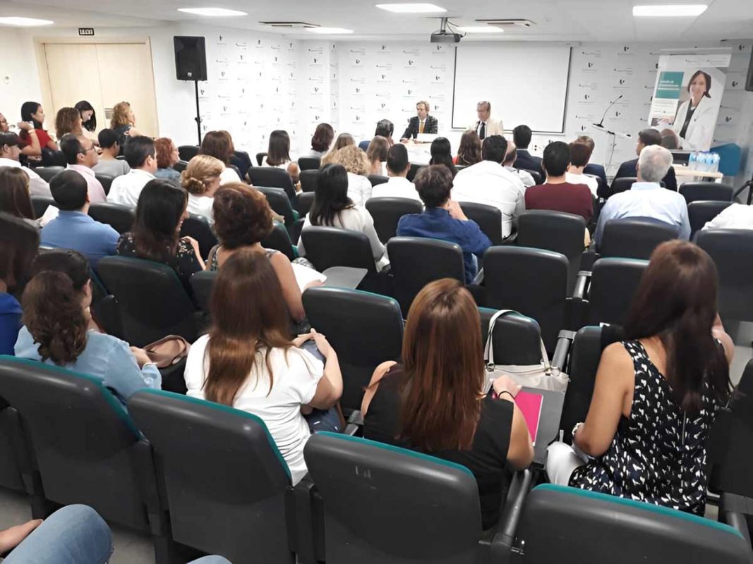
Quirónsalud Málaga Hospital celebrated the First Quirónsalud Update Conference on Diagnostic Imaging on 5 October, a multidisciplinary meeting that included presentations on all diagnostic modalities: ultrasound, mammography, MRI and CT scans. The Conference, organised by Quirónsalud Málaga and Quirónsalud Sagrat Cor Hospitals, brought together 90 attendees, including radiologists from all eight Andalusian provinces, as well as the heads of all Quirónsalud radiology departments in Andalusia.
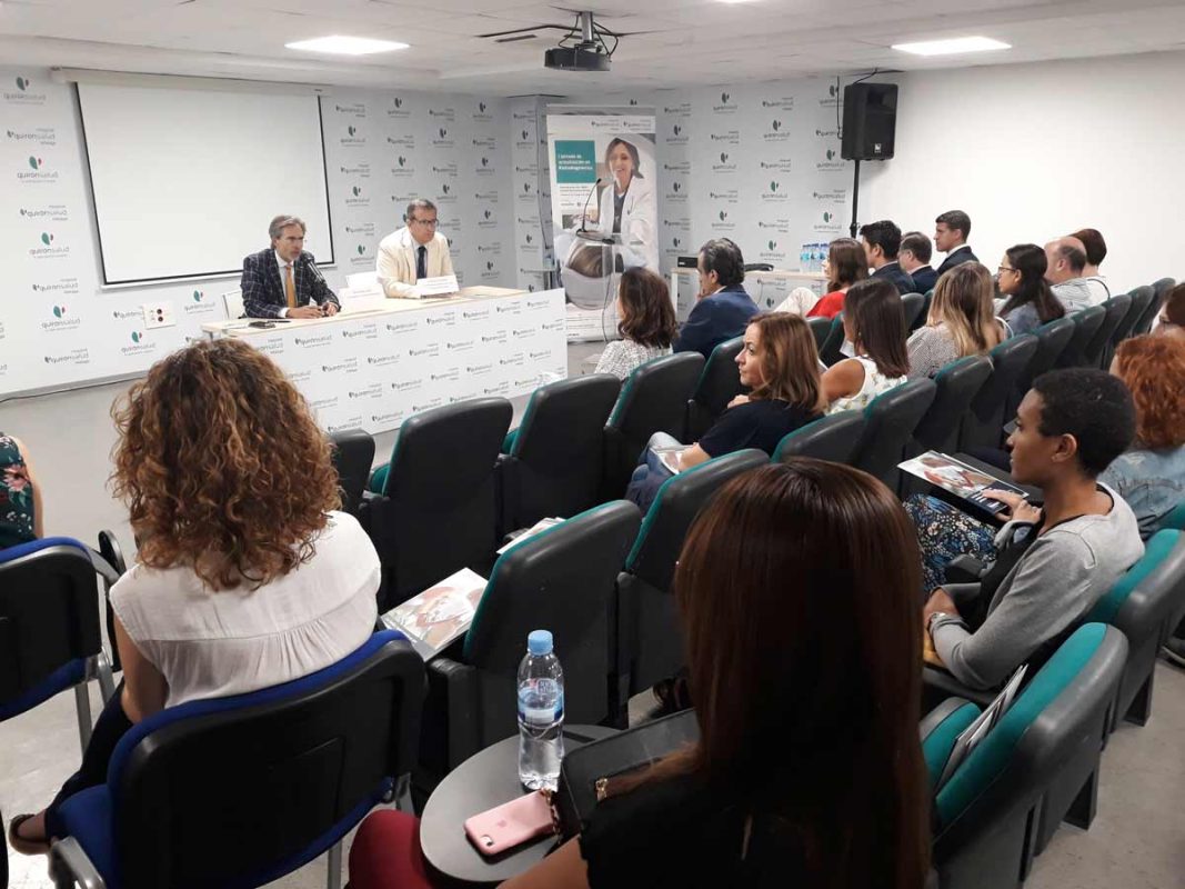
The meeting was presented by Dr. Tomàs Urda, CEO of Quirónsalud Hospitals Malaga and Marbella; and Dr. Carlos Alonso, Head of the Radiology and Medical Imaging Service at Quirónsalud Hospital Malaga. Among the speakers, the presence of Dr. Pablo Valdés, President of the Spanish Society of Medical Radiology, and Dr. Joan Carles Vilanova, Medical Director of the Magnetic Resonance Service at Clínica Girona and President of the latest scientific committee of this year's national congress, is noteworthy.
In the words of Dr. Carlos Alonso, Head of Radiology at Quirónsalud Málaga and organiser of the conference, “this meeting aims to be, today and in future editions, a meeting point for Andalusian radiologists, where they can discuss the latest topics, including the implementation of tomosynthesis or multiparametric prostate MRI.“.
Dr Ignacio Galán, from the Quirónsalud Sagrat Cor Hospital, gave the first presentation, which focused on the present and future of breast tomosynthesis. “Tomosynthesis creates a 3D image of the breast, which increases the sensitivity and specificity of mammography, significantly reducing indications for unnecessary biopsies thanks to this technique. Recent studies have shown a% 35% increase in the breast cancer detection rate thanks to the use of this technology,“ he points out.
AI to improve cancer detection
The multiparametric MRI of the prostate was the next topic to be discussed, with Dr Joan Carles Vilanova, from Clínica Girona-IDI, who commented that “this technique can reduce overdiagnosis and improve cancer detection”.
Subsequently, Doctor Ernesto Rivera, who leads the Unit for Percutaneous Treatments of Musculoskeletal Lesions at Quirónsalud Málaga Hospital, elaborated on arthro-magnetic resonance imaging (Arthro-MRI) of the upper limb. “By introducing contrast into the joint and distending it before the resonance, Arthro-MRI substantially improves the assessment of joint lesions, many of which are not detected by other imaging techniques.”.
Dr Pablo Valdés, from the Costa del Sol Hospital, defended the future of Radiology in Spain, pointing out that “new radiological technologies could increase efficiency and the capacity for detection, characterisation and staging of pathologies by being able to combine different imaging techniques.“.
Meanwhile, Norberto Villajos, a Magnetic Resonance specialist from General Electric Healthcare, presented advances in CT and MRI. In parallel, Doctor Gemma Pérez, from Quirónsalud Camp de Gibraltar Hospital, explained the management of thyroid nodules. “The incidence of cancers smaller than 1 cm has increased by 49% and those smaller than 2 cm by 87%, which are diagnosed thanks to the improvement in imaging methods,’ she commented.
Dr Joan Carles Vilanova then took the floor again to speak about whole-body MRI. He was followed by Dr Ana Carvajal, from Quirónsalud Málaga, who analysed pelvic floor pathology with MRI, pointing out that “MRI is a useful and easily executable technique in those cases where clinical examination is not sufficient for a convenient diagnostic assessment“.
The following presentation was given by Dr Iván Artero, from Quirónsalud Marbella Hospital, who described the appropriate use of teleradiology and its utility in the diagnostic imaging service. Afterwards, Dr Juan Mesa, from Quirónsalud Córdoba Hospital, reviewed the current indications for contrast digestive studies.
To conclude the day, Dr. Esther García-Valdecasas, from Quirónsalud Sagrat Cor Hospital, gave a presentation on pulmonary arteries and veins with MDCT. “Multidetector Computed Tomography (MDCT) is a very useful diagnostic tool that allows us to understand the anatomy of the pulmonary veins, their anatomical variations, and the existence of anomalous venous drainage, which is impossible with other imaging techniques.”.